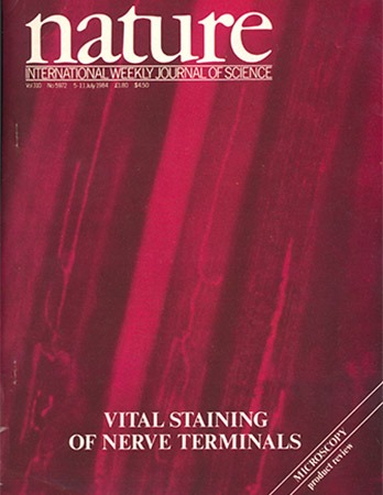
Biology Under Cover
Selected Journal & Book Covers from SBS Faculty
"Staining of living presynaptic nerve terminals with selective fluorescent dyes"
Doju Yoshikami, Lawrence Oken
Summary
Johnson et al. have recently shown that several positively charged, membrane-permeant fluorescent dyes can serve as vital stains for mitochondria in cultured cells1,2 (see also ref. 3). We report here that presynaptic nerve terminals, which are characteristically rich in mitochondria4,5, can also be vitally stained with such dyes, and that this staining offers a resolution of structural detail not available with previous methods for visualizing terminals in living tissues. The dyes have provided excellent, highly detailed fluorescence images of presynaptic motor nerve terminals at neuromuscular junctions in conventional live tissue preparations from mouse, frog and Drosophila. In addition, when tested at the frog neuromuscular junction, at least one of the dyes permitted visualization of terminals with little, if any, effect on synaptic transmission. Moreover, we have found that by using the dyes it is now also possible to see motor nerve terminals in situ in live animals.
